


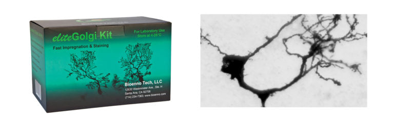
eliteGolgi Kit (Cat#006690)
eliteGolgi Kit (Catalog Number 006690)
(Distributor and quantity discounts available, please send an email to
contact@bioenno.com to order.
For common questions and answers, please check our FAQ.)
Details and FeaturesFast Impregnation and Staining, Excellent for Fresh Tissues
The eliteGolgi Kit (Cat # 006690) is ideal for staining neurons in the olfactory bulb, cortex, cerebellum, brain stem, and spinal cord. The kit supplements and extends the capability of our proven superGolgi Kit (Cat # 003010). This kit can be used in combination with the superGolgi Kit and sliceGolgi Kit (Cat # 003760) to achieve a more comprehensive analysis of select areas of the brain (see Table below — The table is created as a reference for users to determine which Golgi Kit would be the best for staining neurons in specific regions of a mouse/rat brain).
Table: Staining Selectivity of the Bioenno Golgi Kits
|
Region |
Bioenno Golgi Kits |
||
|
superGolgi Kit (# 003010) |
eliteGolgi Kit (# 006690) |
sliceGolgi Kit (# 003760) |
|
|
Olfactory bulb |
++ |
+++ |
++ |
|
Cortex (e.g., Frontal, Piriform, Entorhinal, Perirhinal, and Neo) |
+++ |
+++ |
+++ |
|
Hippocampal formation |
+++ |
-~+ |
++ |
|
Amygdala |
+++ |
-~+ |
++ |
|
Striatum |
++ |
- |
+++ |
|
Septum |
+++ |
+ |
++ |
|
Nuclei in brainstem |
+ |
+++ |
+~++ |
|
Cerebellum |
- |
+++ |
-~+ |
|
Spinal cord |
+~++ |
+++ (immature rats/mice) |
+~++ |
The eliteGolgi Kit has been rigorously tested and validated on brain tissues freshly harvested from rats and mice (see Proven Results below). This kit allows neurons to be impregnated rapidly requiring only 3-6 days depending on the age, size, and region of the tissues. Moreover, the staining of neurons can be performed on either free-floating or mounted sections (50~250 µm thickness). The kit can be stored in a dark area at room temperature (4-25ºC) for up to 12 months.
Important! The eliteGolgi Kit does not work well on frozen tissues.
Product Features
- Reliable and high-contrast staining of cells in the superficial regions of the brain
- Suitable for freshly harvested brain tissues (Not for 4% PFA-fixed, frozen tissues)
- Impregnation time of only 3-6 days
- Streamlined staining protocol
- Sufficient for up to 50 mouse brains
- For in-vitro lab use
- Warranty: 12 months
- EliteGolgi Kit Description and Protocol.
- Sample is available upon request. Test Sample Description and Protocol.
In a number of investigations, neurons and dendritic spines have been reliably stained with the eliteGolgi Kit. The following shows some representative images.
Fig. 1 – Golgi impregnated and stained neurons in the neocortex
The eliteGolgi Kit was used to impregnate and stain the neurons in the neocortex of a postnatal day 18 Sprague Dawley rat. The boxed area in the left panel (taken at 20x objective lens) was magnified (63x) to show the dendrites, axon, and soma of a stained neuron.

Fig. 2 – Impregnated and stained cells in the olfactory bulb and spinal cord
The stained cells in the olfactory bulb (left panel, taken at 10x objective) and the spinal cord (right, 4x objective) of a postnatal day 11 rat. Freshly harvested tissue blocks were subjected to Golgi staining via the eliteGolgi Kit. The impregnation time was 4 days at room temperature (22 ± 1ºC). The sections were counterstained with Bioenno Cresyl Violet (Cat # 003003).

Fig. 3 – Impregnated and stained cells in the olfactory bulb (sagittal section)
The eliteGolgi Kit was used to impregnate and stain the cells in the olfactory bulb of a 12-day-old Sprague Dawley rat. The boxed area in the left panel was magnified (63x) to show the branches of a stained cell.

Fig. 4 – Golgi impregnated and stained cells in the spinal cord (wet section)
Golgi staining was performed on saline-perfused spinal cord tissue block (12-day-old rat) using the eliteGolgi Kit. The impregnation time was 4 days (22 ± 1ºC). These images were taken from a wet section (not dehydrated in ethanol, not cleared in xylene or xylene substitute) using 2x, 10x, or 20x objective lenses. The boxed areas were magnified (10x or 20x objective) to show cells in the posterior horn of the spinal cord.

Fig. 5 – Golgi impregnated and stained cells in the medulla (dark field images)
Golgi staining was performed on saline-perfused brain stem tissue block using the eliteGolgi Kit. The dark field images were taken from a wet section (not dehydrated in ethanol, not cleared in xylene or xylene substitute) of 12-day-old rat. The area of pyramidal tract was dotted. Cells in the boxed area was magnified (20x) to show the pattern of their dendritic branches.

Fig. 6 – Golgi impregnated and stained Purkinje cells in the cerebellum (wet section)
Golgi staining was performed on freshly harvested cerebellum (not PFA-fixed) of a 12-month-old C57 mouse using the eliteGolgi Kit. The impregnation time was 4 days (22 ± 1ºC). The image was taken from a wet section (not dehydrated & not cleared) using a 20x objective lens.

Fig. 7 – Golgi-stained dendritic branches of Purkinje cells
The eliteGolgi Kit was used to impregnate and stain the dendritic branches of Purkinje cells in the cerebellum of a 6-month-old C57 mouse. The impregnation took 4 days only. The staining and clarity took 3 min and ~1 min, respectively. The section was air-dried, cleared in xylene, and cover slipped with Permount® mounting medium.

Fig. 8 – Neurons in the anterior olfactory nucleus
The eliteGolgi Kit was used to impregnate and stain the neurons located in the anterior olfactory nucleus (lateral) (left panel, 4x objective) of a 12-day-old rat. The boxed area was magnified (20x) to show a stained neuron and further magnified (63x) to show the dendritic spines.

Fig. 9 – Neuron in the frontal cortex
The eliteGolgi Kit was used to impregnate and stain cells in the frontal cortex (left panel, 4x objective). The framed area was magnified (20x) in the right to show the branches and dendritic spines of a pyramidal neuron located in layer III.

Fig. 10 – Golgi stained neurons in layer II of the frontoparietal cortex
The stained cells in the frontoparietal cortex (left, taken at 20x objective) of a postnatal day 18 rat. The framed area was magnified (100x objective) in the right to show the dendritic spines. Freshly harvested tissue blocks were subjected to Golgi staining via the eliteGolgi Kit. The impregnation time was 3 days only (22 ± 1ºC). The section was counterstained with Bioenno Cresyl Violet (Cat # 003003).
Fig. 11 – Two neurons in the somatosensory cortex
The eliteGolgi Kit was used to impregnate (4 days, 22 ± 1ºC) and stain (3 minutes, 22 ± 1ºC) neurons in the frontoparietal cortex (somatosensory area). The images were taken from the cortex of a 12-day old rat using a 20x objective lens.
Fig. 12 – The neurons in the spinal trigeminal nucleus
The eliteGolgi Kit was used to impregnate (4 days, 22 ± 1ºC) and stain (3 minutes, 22 ± 1ºC) neurons in the spinal trigeminal nucleus. The image was taken from a 6-month-old C57 mouse using a 10x objective lens.

Fig. 13 – Cells in the lateral hypothalamic area
The eliteGolgi Kit was used to stain cells in the lateral hypothalamic area (left, 10x objective) of a 11-day-old rat. The boxed area was magnified (right, 63x objective) to show the filopodia-like protrusions (arrows), the immature spines. The impregnation time was 4 days (22 ± 1ºC) and staining time was ~4 minutes (22 ± 1ºC).
brain tissue, dendrite, dendritic spine, GFP, gfp, golgi cox, golgi impregnation, golgi stain, golgi’s method, impregnation, rapid golgi, spine, staining, super golgi, supergolgi, synapse