


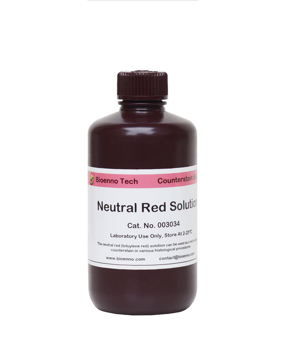
Neutral Red Solution (Cat#003034)
Neutral Red Solution (Catalog Number 003034)
(Laboratory Use Only, Store at 2-25 ºC)
$120 USD/Solution
(Distributor and quantity discounts available, please send an email to
contact@bioenno.com to order.
For common questions and answers, please check our FAQ.)
The Neutral Red (Toluylene Red) Solution can be used as a red nuclear counterstain in various histological procedures. This solution has been proven to generate excellent nuclei staining on frozen tissue sections and paraffin-embedded brain sections with or without enzyme histochemistry and/or immunohistochemistry. The staining can be carried out at room temperature (18-25ºC) and takes approximately 1-5 min. Staining time should be optimized for each tissue type and staining intensity desired. This counterstain solution is suitable for use with non-aqueous mountants.

Neutral Red Solution used together with Bioenno DAB-Co Substrate Kit and superGolgi Kit
(A) Section stained with the Neutral Red solution. (B) Bioenno DAB-Co Kit was employed to develop the immunoreactive neurons (bluish black), followed by Neutral Red counterstaining. (C) Bioenno superGolgi Kit was used to stain the dendritic spines (150 µm thickness of sections), followed by Neutral Red counterstaining. The box in C1 was magnified in C2. All the images were taken from adult mouse brains.
Warranty: 12 months from the date of purchase.
Return Policy: Bioenno Tech’s return policy for this product is 90 days from the date of purchase.
Free Technical Support: Email your questions to contact@bioenno.com
REAGENTS PROVIDED:
- Neutral Red: 250 ml of solution in a plastic bottle. The solution can be used as a red nuclear counterstain in various histological procedures.
Instructions for Use:
(A) Sections with completed immunohistochemistry (IHC/ICC), in situ hybridization (ISH), or Golgi staining:
- Finish the IHC/ICC, ISH, or Golgi staining, and mount the tissue sections upon adhesive microscope slides. Air-dry the slides at room temperature (RT, 18-25ºC).
- Wash the dry slides in 0.01 M PBS-T for 1-5 minutes and then rinse in dH2O for 1-3 seconds.
- Individual Slide Staining: Place the slides on a level surface and apply the solution to sections. Make sure the sections are fully covered with the solution. Incubate the sections at RT for 1-5 minutes depending on the desired intensity and type of tissues. After incubation, wash out extra counterstain solutions with dH2O.
Batch Staining: Put the Neutral Red Solution in a staining jar, and add slides and incubate at RT for 1-5 minutes. After incubation, remove slides and rinse off excess stain solution with dH2O.
- Check the stained sections under microscope for the best results.
- Optimal time should be determined by the investigators. Longer time of incubation may be required for some specific tissues.
- The Solution can be reused. If necessary, filter the solution with 0.2-0.4 µm syringe type filter or WhatmanTMfilter paper before use. To enhance the staining intensity, the solution can be warmed up to 40~45ºC before the staining.
- If needed, differentiate the sections in the kit provided acidic ethanol or 70% ethanol for seconds to minutes, and check microscopically for best result. Wash out the ethanol with dH2O.
- The staining intensity of both cellular elements and background decreases quickly in the acidic ethanol or 70% ethanol.
- If staining is light, simply reapply the counterstain solution and incubate the sections again.
- Air-dry the slides and then directly dehydrate sections in 100% ethanol for 1-2 times, 3-5 minutes each. Clear in xylene or xylene substitute, 2-3 changes, 3-5 minutes each. Cover slip with Permount® mounting medium.
- Longer time of Dehydration and Clear may be required for thick Golgi-stained sections.
(B) Routine histological sections:
- Sections should be mounted on gelatin coated or positive charged plus slides. The paraffin-embedded sections should be dewaxed before running the staining.
- Air-dry the sections/slides. Wash the slides in 0.01 M PBS-T for 1-5 minutes and then rinse in dH2O for 1-3 seconds.
- Stain in the counterstain solution for 1-5 minutes as described above.
- If needed, differentiate in acidic ethanol or 70% ethanol for seconds to minutes and check microscopically for best result.
- Dehydrate in 100% ethanol, clear in xylene or xylene substitute, and cover slip with non-aqueous mountants as described above.
Storage, Safety, and Handling Precautions:
- Store the solution at 2-25ºC and avoid strong direct light.
- The solution is designed for in vitro research use only and not for drug, diagnostic or other uses.
- The solution contains reagents that may be harmful in contact with skin, by inhalation or ingestion. Do not pipette by mouth. Use ordinary precautions to avoid inhalation and contact with skin and eyes. In case of contact, wash immediately with generous amounts of water and seek medical advice. If swallowed, wash out mouth with water and immediately call a physician.
- Perform experiment under a chemical hood. Wear suitable protective clothing, gloves and eye/face protection. Wash hands thoroughly after performing the experiment.
- Material safety data sheet (MSDS) is available upon request.
brain tissue, dendrite, dendritic spine, GFP, gfp, golgi cox, golgi impregnation, golgi stain, golgi’s method, impregnation, rapid golgi, spine, staining, super golgi, supergolgi, synapse