


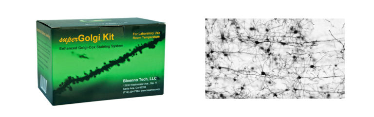
superGolgi Kit (Cat#003010)
An Enhanced Golgi-Cox Impregnation & Staining System For Dendrites and Spines.
superGolgi Kit Product Brochure
USD $483 / kit
(Distributor and quantity discounts available, please send an email to
contact@bioenno.com to order.
For common questions and answers, please check our FAQ.)
The Golgi Method and superGolgi Kit – An Overview
The superGolgi Kit provides an enhanced Golgi-Cox staining system. The superGolgi Kit is designed for the staining and impregnation of dendrites and dendritic spines of neurons. It is based on the principle of Golgi-Cox impregnation. The Kit has been extensively tested on various brain tissues including those harvested from rats, mice, cats, rabbits, monkeys, as well as postmortem human brains. See The Golgi Method and superGolgi Kit – An Overview and Introduction to the Golgi Method for an overview about this technique and its uses in the neurohistology field.
The superGolgi Kit yields both stable and high quality labeling of dendrites and spines. The impregnation time takes 7 to 14 days depending on the age and size of tissues. The Kit can be stored in a dark area at room temperature (22 +/-1℃) for up to 18 months.
Product Features
- Reliable and high contrast impregnation and staining of dendrites and dendritic spines
- Suitable for freshly harvested brain tissues
- Extensively tested on a broad range of species
- Employs a refined and stable Golgi-Cox solution
- Impregnation time of 1 to 2 weeks
- Streamlined staining protocol
- Sufficient for 10-12 blocks (~1 x 1 x 2 cm) of brain tissue
- For in-vitro lab use
- Warranty: 18 months
Product Description and Protocol
Proven Results
Dendritic branches and spines have been reliably stained and impregnated with the superGolgi Kit.

Fig. 1 – Impregnated and Stained Brain Sections Mounted on Adhesive Glass Slides
As shown in Fig. 1, following the superGolgi Kit protocol, brain tissues from a rat and mouse were impregnated, sliced, and stained. The stained sections can be stored at room temperature.

Fig. 2 – Impregnated and Stained Pyramidal Neurons in the Cortex (P2)
As shown in Fig. 2, the superGolgi Kit was used to impregnate and stain the pyramidal neurons in the neocortex of a postnatal day 2 (P2), C57BL mouse (20x objective lens). Dendrites and axons were well impregnated and stained.

Fig. 3 – Pyramidal Neuron in the Hippocampal CA1 Area (P2)
As shown in Fig. 3, the superGolgi Kit was used to impregnate and stain the pyramidal neuron in the CA1 area of the hippocampus of a P2 C57BL mouse (20x objective).

Fig. 4 – Pyramidal Neuron in the Hippocampal CA3 Area (P7)
As shown in Fig. 4, the superGolgi Kit was used to impregnate and stain the pyramidal neuron in the CA3 area of the hippocampus of a P7 C57BL mouse (20x objective). The boxed area was magnified to show the filopodia-like protrusions (arrowheads), which are the immature dendritic spines.

Fig. 5 – Pyramidal Neurons and Labeled Dendritic Protrusions(P21)
As shown in Fig. 5, pyramidal neurons were stained using the superGolgi Kit. The pyramidal neurons were taken from the frontoparietal cortex (motor area) of a P21 CD1 mouse. The boxed areas in the Middle panel, shown at 20x, were magnified to 100x and presented in the Left and Right panels. On the Left panel, oblique branches are shown. Arrowhead-indicated, filopodia-like protrusions, which are the immature dendritic spines, are often observed at this age. On the Right panel, mature dendritic spines on the main branch are indicated by arrows.

Fig. 6 – Impregnated Cortical Neurons and Labeled Dendritic Protrusions (P30)
As shown in Fig. 6, cortical neurons were stained using the superGolgi Kit. These neurons were taken from the frontoparietal cortex (somatosensory area) of a P30 C57BL mouse. The boxed area in the Top panel, shown at 10x, was magnified (63x objective) to highlight the dendritic protrusions.

Fig. 7, Left and Right – A Striatal Neuron and Labeled Dendritic Spines (denoted by arrows) (2-month)
As shown in Fig. 7, the superGolgi Kit was used to impregnate and stain the striatal neuron which was taken from the posterior caudate of a 2-month old Wistar rat. (Left Panel: 20x objective; Right Panel: Boxed area was magnified to 63x)

Fig. 8 – Impregnated and Stained Hippocampus (5-month)
As shown in Fig. 8, the superGolgi Kit was used to impregnate and stain the hippocampal neurons of a 5-month old C57BL mouse (4x objective lens). “DG” denotes dentate gyrus.
Importance of Golgi-Cox Impregnation and Staining
Golgi-Cox impregnation and staining allows scientists to clearly visualize the soma, dendrites, and dendritic spines of neurons from the brain tissues of various animals including rats, mice, cats, rabbits, and monkeys, as well as tissues obtained from post-mortem human brains. Understanding the morphology of these cellular structures is critical for research on the effects of various diseases on the brain including Alzheimer’s. In addition, understanding these structures allows scientists to better understand the effects of aging and the effects of man-made toxins and illicit drugs (e.g. methamphetamine) on the brain. Furthermore, Golgi-Cox impregnation and staining allows scientists to further understand neuroplasticity and neuroprotection.
Importance of Dendrites of Neurons
Dendrites of neurons make up 95% of the total volume of the neuron. Synapses, which are structures that permit neurons to pass electrical and chemical signals to another cell, are found on dendrites. If there are any changes to the dendrites, this will cause neuron damage. It is important to characterize these dendrites to determine if there is any branching atrophy or spine loss.
FAQ about the superGolgi Kit
· I am using the superGolgi Kit. Is it necessary to perfuse the animal with saline? What’s the consequence of staining without perfusion?
It is recommended to perfuse the animals with saline. The perfused brains will quickly be well impregnated compared to non-perfused samples. The background is also better in the perfused samples vs. non-perfused samples. However, there is no observed difference in the staining of dendritic spines.
· I am using the superGolgi Kit. The impregnation is complete and the brains are ready for slicing. However, I have no time to cut these brains. How do I store these impregnated brains?
The impregnated tissue blocks can be stored in the post-impregnation solution for up to 4 weeks at 4ºC. Once you initiate the cutting, please finish the cutting, staining, and all following steps ASAP.
· I have no access to a vibratome, but do have access to a cryostat. How do I use the cryostat to cut impregnated brain tissue blocks?
It is recommended to slice the impregnated tissues under room temperature (20~22ºC). We recommend the usage of a vibratome or micro slicer as the aforementioned units will expedite the cutting process.
A cryostat may be used, but not under usual operation – this means the temperature must be set to 20~22ºC, or simply power off the machine and then treat the cryostat as a micro slicer. Glue the tissue block onto the chuck by using Krazy Glue or Super Glue (No freezing and No embedding).
A problem for cutting thick sections such as 100, 200, 300 micron is that the designed thickness for most cryostats is 1-60 micron. So how do I cut a 120 um section for example? Our experience is to set the thickness at 60 um, turn the slicing knob clockwise almost half-circle. Then turn the knob counterclockwise BACK to the starting position. By continuing the process this will yield one 120 micron section.
[Note: This method works on our machines, but it is not a universally compatible technique. Using this method may cause damage to your specific machine depending on make and model. Be advised this is a demonstration of one methodology to serve as a reference. ]
· I am using the superGolgi Kit. Can I perform the staining on free-floating sections?
Yes, the staining and post-staining can be performed on free-floating sections to save the reaction solutions C and D, but the mounting of sections may take a while.
It is recommended to perform the staining on mounted sections because the impregnated tissues/sections are frangible. To prevent sections from breaking during the collection/transfer process, a large shader paintbrush is provided with the kit.
· I am using the superGolgi Kit. How do I prevent sections from falling off the slide during staining and post-staining?
The sections should be mounted on adhesive microscope slides, such as gelatin-coated slides, and you can gently press the sections upon the slide using bibulous paper. Wear gloves to avoid any oil or lotion from hands when mounting the sections. Air-dry the sections before washing in PBS-T and avoid washing of sections/slides in dH2O.
Some publications cited with superGolgi Kit
(For more detail, please see Publication List)
-
5-Fluorouracil chemotherapy upregulates cytokines and alters hippocampal dendritic complexity in aged mice
Thomas R. Grovesa, Ryan Farrisd, Julie E. Andersona, Tyler C. Alexandera, Frederico Kiffera, Gwendolyn Cartera, Jing Wanga, Marjan Boermaa, Antiño R. Allen(2017), Behavioural Brain Research,316, 215–224
Link: http://www.sciencedirect.com/science/article/pii/S0166432816305575
-
Neuropsin Inactivation Has Protective Effects against Depressive-Like Behaviours and Memory Impairment Induced by Chronic Stress
Simon Chang, Philane Bok, Cheng-Pu Sun, Andrew Edwards, Guo-Jen Huang(2016), PLoS Genet, 12 (10), e1006356
Link:http://journals.plos.org/plosgenetics/article?id=10.1371/journal.pgen.1006356 -
Contrasting the effects of proton irradiation on dendritic complexity of subiculum neurons in wild type and MCAT mice
Nicole N. Chmielewski, Chongshan Caressi, Erich Giedzinski, Vipan K. Parihar, Charles L. Limoli(2016), Environmental and Molecular Mutagenesis, 57, 364-371
Link:http://onlinelibrary.wiley.com/doi/10.1002/em.22006/full -
Postnatal Gene Therapy Improves Spatial Learning Despite the Presence of Neuronal Ectopia in a Model of Neuronal Migration Disorder
Huaiyu Hu, Yu Liu, Kevin Bampoe, Yonglin He and Miao Yu(2016), Genes, 7(12), 105
Link:http://www.mdpi.com/2073-4425/7/12/105 -
Adult microbiota-deficient mice have distinct dendritic morphological changes: differential effects in the amygdala and hippocampus
Pauline Luczynski, Sean O. Whelan, Colette O’Sullivan, Gerard Clarke, Fergus Shanahan, Timothy G. Dinan and John F. Cryan(2016), European Journal of Neuroscience, 44, 2654–2666
Link: http://onlinelibrary.wiley.com/doi/10.1111/ejn.13291/pdf -
Cranial grafting of stem cell-derived microvesicles improves cognition and reduces neuropathology in the irradiated brain
Janet E. Baulcha, Munjal M. Acharyaa, Barrett D. Allena , Ning Rua , Nicole N. Chmielewskia , Vahan Martirosiana , Erich Giedzinskia , Amber Syagea , Audrey L. Parka , Sarah N. Benkea , Vipan K. Parihara , and Charles L. Limolia (2016). PNAS, 113 (17), 4836–4841.
Link: http://www.pnas.org/content/113/17/4836.full -
RBM4 promotes neuronal differentiation and neurite outgrowth by modulating Numb isoform expression
Woan-Yuh Tarna, Hung-Che Kuoa, Hsin-I. Yua, Shin-Wu Liua, Ching-Tzu Tsenga, Dodda Dhananjayaa, Kuan-Yang Hunga, Chi-Chiang Tua, Shuo-Hsiu Changa, Guo-Jen Huangb, and Ing-Ming Chiuc (2016). Molecular Biology of the Cell, 27(10),1676-83.
Link: http://www.ncbi.nlm.nih.gov/pubmed/27009199 -
Combined Treatment With Environmental Enrichment and (-)-Epigallocatechin-3-Gallate Ameliorates Learning Deficits and Hippocampal Alterations in a Mouse Model of Down Syndrome
Silvina Catuara-Solarz, Jose Espinosa-Carrasco, Ionas Erb, Klaus Langohr, Juan Ramon Gonzalez, Cedric Notredame and Mara Dierssen(2016). eNeuro, 3(5), 0103-16.
Link: http://eneuro.org/content/3/5/ENEURO.0103-16.2016
-
Targeted overexpression of mitochondrial catalase prevents radiation-induced cognitive dysfunction
Parihar, V. K., Allen, B. D., Tran, K. K., Chmielewski, N. N., Craver, B. M., Martirosian, V., … & Limoli, C. L. (2015). Antioxidants & Redox Signaling, 22(1), 78-91.
Link: http://www.ncbi.nlm.nih.gov/pubmed/24949841 -
Stem Cell Transplantation Reverses Chemotherapy-Induced Cognitive Dysfunction
Munjal M. Acharya, Vahan Martirosian, Nicole N. Chmielewski, Nevine Hanna, Katherine K. Tran, Alicia C. Liao, Lori-Ann Christie, Vipan K. Parihar, and Charles L. Limoli (2015), Cancer Research, 75, 676-686.
Link: http://www.ncbi.nlm.nih.gov/pubmed/25687405 -
Short-term modern life-like stress exacerbates AB-pathology and synapse loss in 3xTg-AD mice
David Bagliett-Vargas, Yuncai Chen, Dongjin Suh, Rahasoon R. Ager, Carlos J. Rodriguez-Ortiz, Rodrigo Medeiros, Kristoffer Myczek, Kim N. green, Tallie Z. Baram, Frank M. LaFerla (2015). Journal of Neurochemistry, 134, 915-926.
Link:http://www.ncbi.nlm.nih.gov/pubmed/26077803 -
Human endogenous retrovirus-K contributes to motor neuron disease.
Wenxue Li, Myoung-Hwa Lee, Lisa Henderson, Richa Tyagi, Muzna Bachani, Joseph Steiner, Emilie Campanac, Dax A. Hoffman, Gloria von Geldern, Kory Johnson, Dragan Maric, H. Douglas Morris, Margaret Lentz, Katherine Pak, Andrew Mammen, Lyle Ostrow, Jeffrey Rothstein and Avindra Nath (2015). Science Transitional Medicine, 7, 307, 307ra153.
Link:http://stm.sciencemag.org/content/7/307/307ra153.full-text.pdf+html
-
Synergistic effects of amyloid-beta and wild-type human tau on dendritic spine loss in a floxed double transgenic model of Alzheimer’s disease.
Chabrier, M. A., Cheng, D., Castello, N. A., Green, K. N., & LaFerla, F. M. (2014). Neurobiology of Disease, 64, 107-117.
Link: http://www.ncbi.nlm.nih.gov/pubmed/24440055
brain tissue, dendrite, dendritic spine, GFP, gfp, golgi cox, golgi impregnation, golgi stain, golgi’s method, impregnation, rapid golgi, spine, staining, super golgi, supergolgi, synapse